
Prone OLIF with Navigation with Robotics
18 August 2026 · Robotic
Presentation
A man in his 80s presented with worsening low back pain and severe pain in the right leg and thigh. He had undergone two previous spinal operations: removal of a facet joint cyst on the right at L4/5 in 2019, followed by a decompression and posterior spinal fusion from L3 to L5 in 2020.
His medical history included a previous heart attack (myocardial infarction) and severe aortic stenosis — a significant narrowing of one of the main heart valves.
The challenge
Imaging showed that the level immediately above his previous fusion had worn out — a well-recognised problem called adjacent segment disease. At L2/3 there was:
- Collapse of the intervertebral disc
- Retrolisthesis — a backward slip of L2 on L3
- Overgrowth (hypertrophy) of the facet joints on both sides, compressing the nerve roots exiting and passing at the L2/3 level
His heart condition meant he carried a high anaesthetic risk, so the operation needed to be as short and as gentle as possible. A conventional open revision — re-opening the previous midline wound, removing scar tissue and extending the fusion through a large exposure — would have meant a longer anaesthetic and more blood loss than was safe for him.
One alternative was to perform the interbody fusion with the patient lying on his side, then turn and re-drape him to complete the posterior fusion and decompression. That repositioning step adds operative and anaesthetic time, so it was rejected in favour of a single-position technique.
The approach
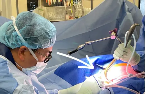
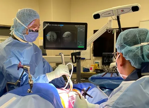
The plan was a prone OLIF (oblique lumbar interbody fusion) at L2/3 with extension of the existing fusion, combined with UBE (unilateral biportal endoscopy) decompression — all performed in a single prone position with robotic navigation.
- Through an incision of about 4 cm, a retroperitoneal window was developed along the anterior-to-psoas (ATP) corridor to reach the L2/3 disc without entering the abdominal cavity.
- The collapsed disc was carefully prepared, using sequential, gentle distraction to protect the fragile endplates of an older spine.
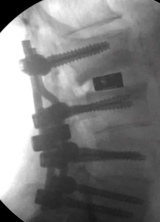
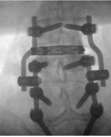
- An expandable cage was placed across the disc space, giving broad coverage of the endplates and restoring disc height.
- The nerve roots were then directly decompressed at L2/3 using UBE — a keyhole endoscopic technique performed through two small portals.
- The fusion was extended by connecting new fixation to the existing implants with connectors and rods, avoiding removal of the previous construct.
Because the whole operation was done in one prone position, there was no need to reposition or re-drape the patient partway through, which shortened the operative and anaesthetic time. Navigated instruments were tracked continuously on the navigation system, allowing accurate placement of implants with minimal radiation exposure.
Imaging and result
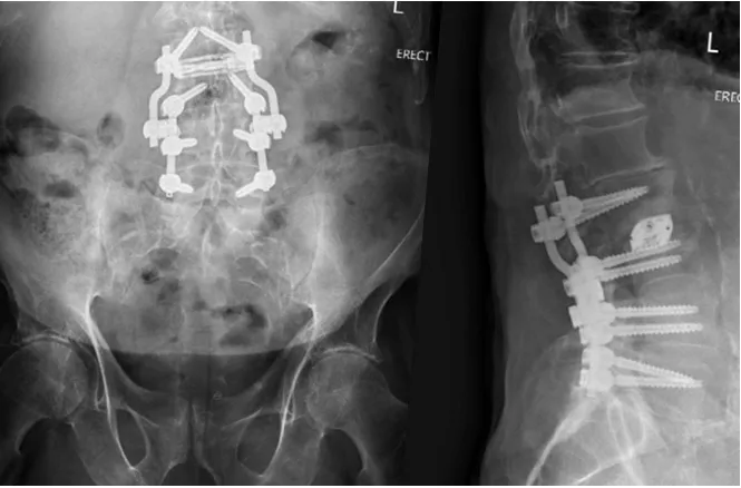
Intraoperative fluoroscopy confirmed placement of the expandable cage in the middle of the disc space, restoration of disc height, and secure connection of the new fixation to the existing implants. Standing X-rays after surgery showed the completed construct with spinal alignment restored. The figures below show the operating theatre set-up and the imaging.
This case study is provided for education only and describes a single patient's course. Individual results vary — outcomes described here do not predict the results of treatment for any other patient.
We are here to listen. We understand your pain. Let us help.
Our professional team is at your disposal — from non-operative treatments through to advanced surgery.
Book a consultationor call 03 5925 6353 (Mornington) · 03 9989 3373 (Melbourne)