
Endoscopic Discectomy for an Acute Disc Herniation with Leg Weakness
19 August 2026 · Endoscopic
Presentation
A man in his early 40s presented with severe low back pain and severe pain down the left leg. The pain began after lifting heavy weights at work, in a job involving regular heavy manual handling.
By the time he was seen he had a four-week history of dense weakness in the left leg. On examination there were both sensory and motor changes in that leg, with power graded 3+ out of 5 on the MRC scale — meaning he could move the leg against gravity and some resistance, but not with normal strength.
Straight-leg raise was positive and tension signs were positive: lifting the straight leg reproduced his pain, which indicates that the sciatic nerve is being stretched over something that is compressing it.
He was otherwise well. Before referral he had already had two nerve-root blocks arranged through his GP, and had not responded to conservative management.
The problem
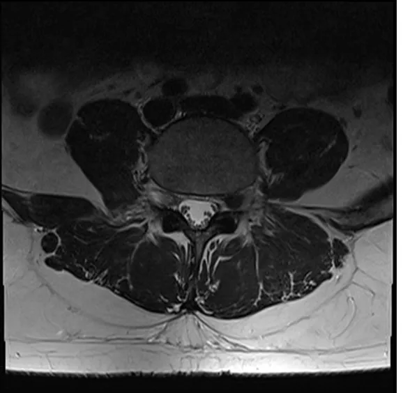
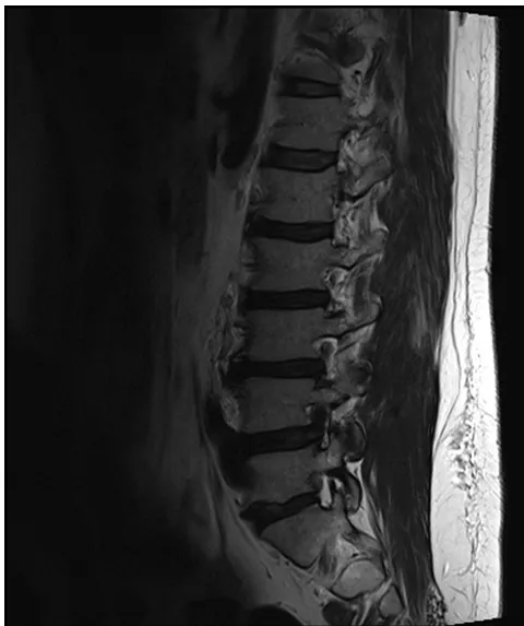
MRI showed a large disc herniation in the lower lumbar spine. A fragment of the disc had extruded backwards into the spinal canal and was compressing the nerve root, which explained both the leg pain and the weakness.
Two features made this more than a pain problem:
- Motor weakness, not just pain. Weakness graded 3+ out of 5 with sensory change indicates that the nerve root is not simply irritated but functionally impaired.
- Time. The weakness had already been present for four weeks. The longer a nerve root remains compressed, the less predictable the recovery of power becomes, so continuing to wait carried a real cost.
Conservative treatment had also been given a fair trial without response, including two nerve-root blocks. Surgery to take the pressure off the nerve was therefore recommended without further delay, with the additional consideration that his work required him to return to heavy lifting.
The approach
The operation was a minimally invasive endoscopic discectomy — removal of the herniated fragment through a small portal under direct endoscopic vision, rather than through an open incision.
- A portal of roughly one centimetre is made over the affected level. The muscle is dilated and worked around rather than stripped off the back of the spine.
- The endoscope gives a magnified, brightly lit view of the nerve root under continuous saline irrigation, so the anatomy is seen close up rather than at a distance through a larger wound.
- A small window is made in the ligament at the back of the spinal canal (the ligamentum flavum) to expose the compressed nerve root.
- The nerve root is gently protected and retracted, and the herniated disc fragment is removed from underneath it, freeing the root.
- Because the bone, the facet joint and the muscle attachments are largely preserved, the segment remains stable and no fusion or implant is required.
The intention of the technique is to relieve the pressure on the nerve while disturbing as little normal anatomy as possible.
Watch the surgical video
The video loads from YouTube only after you open this panel. If it does not load, you can watch it on YouTube. It shows an endoscopic view of the operation and contains surgical footage.
Result
At the end of the procedure the herniated fragment had been removed and the nerve root was decompressed and free.
Recovery after a period of dense weakness follows its own timetable. Pain caused by nerve compression commonly settles once the pressure is removed, but the return of power depends on how long the nerve was compressed and how much it was injured, and is measured over months rather than days. The plan after surgery was therefore a graded rehabilitation program, with strength reassessed at follow-up before any return to heavy manual work.
The MRI images below show the herniation that was treated.
This case study is provided for education only and describes a single patient's course. Individual results vary — outcomes described here do not predict the results of treatment for any other patient.
We are here to listen. We understand your pain. Let us help.
Our professional team is at your disposal — from non-operative treatments through to advanced surgery.
Book a consultationor call 03 5925 6353 (Mornington) · 03 9989 3373 (Melbourne)