
Biportal Endoscopic Decompression and TLIF
19 August 2026 · Endoscopic
Presentation
A man in his late 60s presented with twelve months of low back pain, followed by nine months of progressive pain in the buttock and leg. The right-sided sciatica had been increasing in intensity.
His walking distance had steadily reduced to the point of claudication — leg pain and heaviness brought on by walking and relieved by sitting or bending forward, which is the characteristic pattern of narrowing of the spinal canal.
He had worked through a course of non-operative treatment, including injections with a pain team, without lasting benefit. He was otherwise a well man with no significant medical problems, and he remained an active golfer. He had sought several opinions specifically looking for a minimally invasive option that would let him keep playing.
On examination, straight-leg raise on the right was limited to 40 degrees, and there was weakness in the right L5 nerve root distribution graded 4 out of 5 on the MRC scale.
The problem
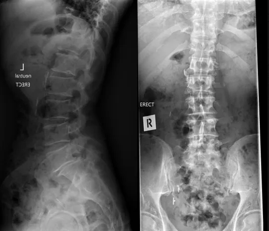
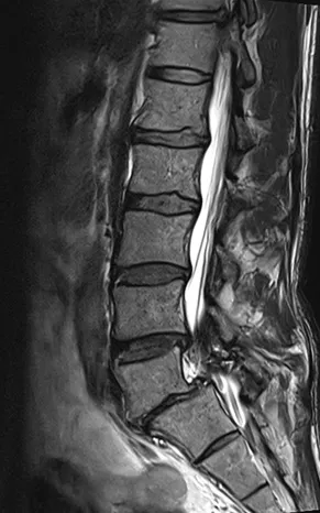
Standing X-rays and MRI showed degenerative change in the lower lumbar spine with narrowing of the spinal canal and compression of the nerve roots. Two levels were responsible, and each needed a different answer:
- L4/5 — the canal was narrowed by thickened ligament and overgrown bone, compressing the nerves as they passed. The segment itself was not unstable, so taking the pressure off was the treatment required.
- L5/S1 — the disc at this level was degenerate as well as compressing the nerve root, and this was the source of both his back pain and the right L5 weakness. Decompression alone would leave a painful, worn-out segment behind, so this level needed to be stabilised as well as decompressed.
Non-operative care had already been given a proper trial. With claudication limiting his walking, sciatica increasing, and measurable weakness in the L5 nerve root, surgery was recommended — and his priority throughout was to have it done in a way that gave him the best chance of getting back onto a golf course.
The approach
Both levels were treated at the same sitting using UBE (unilateral biportal endoscopy) — a keyhole technique in which two small portals are made on the same side of the spine. The endoscope goes through one and the surgical instruments through the other, working under continuous saline irrigation. Because the camera is independent of the instruments, the surgeon can look around corners and work with normal-sized instruments through very small openings.
L4/5 — endoscopic decompression. Under endoscopic vision, the thickened ligament and the overgrown bone pressing on the nerves were removed to reopen the canal. Working from one side, the surgeon can reach across to decompress both sides while leaving the midline structures and much of the facet joint intact. No implant was needed at this level.
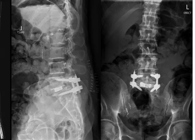
L5/S1 — endoscopic TLIF. Through the same style of portals, the disc was cleared and the endplates prepared, and an interbody cage packed with bone graft was placed into the disc space to restore disc height and support the fusion. Percutaneous pedicle screws and rods were then placed through small stab incisions to hold the segment steady while the fusion consolidates.
Treating both levels endoscopically avoided a single long midline incision and the stripping of muscle off the spine that a conventional open decompression and fusion involves. The muscle is dilated and worked around rather than detached, which is the main reason this approach was chosen for an active patient.
Imaging and result
Standing X-rays before surgery assessed the spine under load, and MRI showed the narrowing of the canal at the lower lumbar levels. Standing X-rays taken after surgery confirmed the interbody cage sitting in the L5/S1 disc space with the pedicle screws and rods in position, and the L4/5 level decompressed without instrumentation, as planned.
Recovery after a two-level procedure of this kind is staged. The plan after surgery was to rebuild walking distance first, with a graded return to golf as the fusion consolidated over the following months. The images below show the pre-operative films and MRI alongside the post-operative X-rays.
This case study is provided for education only and describes a single patient's course. Individual results vary — outcomes described here do not predict the results of treatment for any other patient.
We are here to listen. We understand your pain. Let us help.
Our professional team is at your disposal — from non-operative treatments through to advanced surgery.
Book a consultationor call 03 5925 6353 (Mornington) · 03 9989 3373 (Melbourne)